cryptophyte cortical centrin network
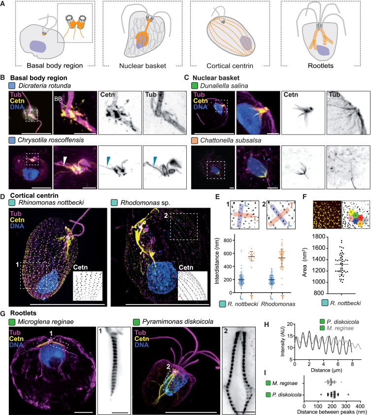
A peripheral centrin-positive cytoskeletal network observed in cryptophyte cells by ultrastructure expansion microscopy. It forms a regular hexagonal mesh in Rhinomonas nottbeckii and shows variable network patterns among Rhodomonas isolates. The assembly is distinct from basal-body-associated centrin structures and from the periplast itself. [DOI:10.1016/j.cell.2025.09.027]
NCBITaxon:1326783) · Felix Mikus and coauthors, Charting the landscape of cytoskeletal diversity in microbial eukaryotes, Cell (2025), Figure 3, DOI 10.1016/j.cell.2025.09.027. Copyright 2025 The Authors; published by Elsevier Inc.; CC BY 4.0. Unmodified publisher image. · CC_BY_4_0 · source: OTHER S0092867425011274 Figure 3 · DOI:10.1016/j.cell.2025.09.027Part of: GO:0005856 — cytoskeleton (no local record)
Components
| Component | Type | Part of / acts on | Grounding | Genes | Stoichiometry | Essential | Role |
|---|---|---|---|---|---|---|---|
| Centrin | Protein | part of | Reviewed label only (reviewed name; no accession asserted) | UNKNOWN | Centrin-family material detected along the peripheral network using the broad-specificity 20H5 antibody after expansion. Immunolocalization supports membership, not an exact paralogue inventory or an assembly requirement.
|
Canonical examples
NCBITaxon:1326783Rhinomonas nottbeckii — RCC6444, also listed as K-1855 in Table S1, was examined by fixed-cell U-ExM. Figure 3D-F and S2 show the cortical network and hexagonal pattern. The source uses Rhinomonas nottbecki, an explicit synonym of the verified NCBI/ENA canonical name Rhinomonas nottbeckii. Table S1 List of Species row 76 identifies RCC6444; Dataset Annotation row 382 marks cortical centrin in one-species interphase material. [DOI:10.1016/j.cell.2025.09.027]NCBITaxon:37096Rhodomonas — Genus-level identification of cultured BMCC67 and RCC10777 (other code Q1-11), not an assertion for every species in the genus. Table S1 List of Species rows 69 and 73 identifies the cultures; Dataset Annotation rows 402 and 412 mark cortical centrin in one-species interphase material. Figure 3D-E and S2 illustrate Rhodomonas networks, but an exact figure-panel-to-isolate mapping was not established. [DOI:10.1016/j.cell.2025.09.027]
Discussions
- CURATION_TODO (RESOLVED): Distinguish this cortical network from centrin proteins and other centrin assemblies. Shared antibody reactivity and superficial mesh geometry do not establish identity or homology between different subcellular assemblies.
- CURATION_TODO (OPEN): Resolve an exact source-neutral centrin family and the network paralogues. Broad antibody specificity cannot identify every gene product, and a broader calcium-binding family must not be promoted to exact centrin identity.
- CURATION_TODO (OPEN): Keep neighboring labeled structures separate and resolve the network-periplast relationship. Colocalization within a whole-cell figure does not make every signal a network constituent.
- CURATION_TODO (OPEN): Determine network diversity and prevalence using comparable labeling across cultures. Sampling and antigen detection constrain distribution claims, while geometry observed in one culture need not define every cryptophyte network.
- CURATION_TODO (OPEN): Test contraction, calcium responses, mechanical roles and assembly dynamics. Fixed-cell morphology and generic centrin biology do not demonstrate a network-level physiological mechanism.
- CURATION_TODO (OPEN): Reconcile expansion correction before recording native dimensions. Conflicting scale statements must not be silently converted into biological measurements.
- CURATION_TODO (RESOLVED): Preserve canonical taxon names and culture-level limits without guessing panel assignments. Culture codes, source spellings and genus-level identifications require explicit reconciliation.
Datasets
- Microbial eukaryote U-ExM volumes and annotations · OTHER · STRUCTURAL_IMAGING · Platform: Ultrastructure expansion microscopy with post-expansion immunofluorescence
Read the pinned data/project.json and data/volumes/dataset.json metadata; volumes is the default dataset. Table S1 identifies cortical-centrin-positive examples rcc6444_pfa_cetn-tub-dna_20230704_cl_s00, bmcc67_pfa_nhs-cetn-tub-dna_20240205_fm_01 and rcc10777_pfa_cetn-tub-dna_20230704_cl_s00. No raw volume is hosted or claimed to have been reanalyzed here, and no exact raw-volume-to-figure mapping is asserted.
Evidence
DOI:10.1016/j.cell.2025.09.027Mikus et al. (2025), Charting the landscape of cytoskeletal diversity in microbial eukaryotes, Cell. Read centrin Results, Figure 3 and S2 legends, fixation, U-ExM, microscopy and image-analysis Methods. The authors distinguish the cryptophyte cortical network from ciliate basal-body-associated networks and describe variable mesh morphology, not a demonstrated physiological function.https://www.dudinlab.com/s/Table-S1.xlsxAuthor-linked Table S1 read as structured workbook data. List of Species rows 69, 73 and 76 identify BMCC67, RCC10777 and RCC6444. Dataset Annotation rows 402, 412 and 382 respectively mark cortical centrin in column AK. Image annotations are not independent-cell counts or clade prevalence estimates.GO:0005622Verified non-obsolete intracellular anatomical structure; broad is-a parent for the intracellular network without claiming a membrane or single resolved polymer lattice.GO:0005856— cytoskeleton Verified non-obsolete cytoskeleton. This cortical centrin assembly is part of the cellular cytoskeleton, not equivalent to its entire framework.
Curation history
2026-10-03T20:07:17Zcodex · CREATED_RECORD — Scaffolded by scripts/new_record.py2026-10-03T20:07:56Zcodex · EXPANDED_RECORD — Added centrin immunolocalization evidence, verified taxon and culture provenance, ontology and physiological boundaries, imaging dataset metadata, and the inspected CC BY publisher Figure 3.
Source: data/structures/cytoskeleton/cryptophyte_cortical_centrin_network.yaml