sinister fiber
A striated, wedge-shaped fibrous linker connecting a flagellar basal body to a five-microtubule s-type root in Spermatozopsis similis. Its BAp95-containing assembly is distinct from the root microtubules, SF-assemblin fibers, centrin fibers and proximal interbasal plates. [DOI:10.1083/jcb.140.5.1149]
Image (not hosted — CC_BY_NC_SA_4_0): PMC2132688.1 Figure 7
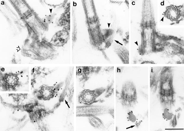
NCBITaxon:3192) · Stefan Geimer, Judith Clees, Michael Melkonian and Karl-Ferdinand Lechtreck, Figure 7, Journal of Cell Biology 140 (1998), doi:10.1083/jcb.140.5.1149, via PubMed Central; CC BY-NC-SA 4.0. Link only; no repository copy or adaptation. · CC_BY_NC_SA_4_0 · source: PMC PMC2132688.1 Figure 7 · DOI:10.1083/jcb.140.5.1149Synonyms: sf-fiber; 1sf-fiber; 2sf-fiber
Part of: GO:0005856 — cytoskeleton (no local record)
Components
| Component | Type | Part of / acts on | Grounding | Genes | Stoichiometry | Essential | Role |
|---|---|---|---|---|---|---|---|
| BAp95 sf-fiber structural proteins | Protein | part of | Reviewed label only (reviewed name; no accession asserted) | UNKNOWN | Fiber-localized structural constituent. Recombinant BAp95 forms striated fibers in vitro, supporting a major structural role without establishing the complete native inventory or native assembly sufficiency.
|
Organism-specific protein examples
Named proteins in which each component's role was established, with the organism the evidence comes from. The component above is the taxon-agnostic family; these are the specific entries behind it.
BAp95 sf-fiber structural proteins
| Accession | Protein | Gene | Organism | Entry |
|---|---|---|---|---|
UniProtKB:O49944 | 95 kD basal apparatus-protein | Spermatozopsis similis (NCBITaxon:3192) | UNREVIEWED | |
Role and evidence (2 references)Sequence-backed BAp95 example corresponding to the cloned antigen; not a complete sf-fiber composition or a curated family accession.
| ||||
Canonical examples
NCBITaxon:3192Spermatozopsis similis — Methods use SAG B1.85. The current SAG 1.85 authority record links this paper's AJ001438 sequence and the historical SAG B 1.85 designation in its sequence metadata. Both 1sf on the older basal body and 2sf on the younger basal body were examined; no clade-wide prevalence is asserted. [DOI:10.1083/jcb.140.5.1149]
Functions
- basal-body to s-root linkage — The native fiber physically links a basal body with its five-microtubule s-type root. This structural linkage does not by itself demonstrate control of phototaxis, spindle placement or cleavage-furrow positioning.
DOI:10.1083/jcb.140.5.1149Results and Figures 7-8 resolve the attachments. The larger 1sf is subdistal on the older basal body; 2sf is proximal on the younger one. Retention of 1sf after mechanical disintegration supports firm basal-body attachment.
Mechanism graphs
Recombinant BAp95 self-assembly in vitro (ASSEMBLY)
Purified recombinant BAp95 forms striated fibers during stepwise denaturant removal. This is an experimental constituent-assembly mechanism, not a reconstitution of the complete native basal-body to s-root linker.
| Subject | Predicate | Object | Evidence |
|---|---|---|---|
| Purified recombinant BAp95 | self-assembles in vitro into | In-vitro striated BAp95 fibers |
|
| Stepwise urea removal in the floating-membrane assay | permits formation of | In-vitro striated BAp95 fibers |
|
Discussions
- CURATION_TODO (RESOLVED): Keep sinister fibers distinct from adjacent basal-apparatus assemblies. Shared location or striation does not establish identical structures.
- CURATION_TODO (RESOLVED): Use a local identifier rather than equating sf-fibers with a mother-centriolar appendage. The paper compares basal feet and satellites by resemblance, without establishing exact homology or identity.
- CURATION_TODO (OPEN): Resolve a source-neutral BAp95 family grounding and the remaining native molecular inventory. A cloned antigen and in-vitro self-assembly do not establish all native constituents or their stoichiometry.
- CURATION_TODO (OPEN): Establish native sf-fiber biogenesis, recruitment and BAp95 essentiality. The recombinant assay establishes intrinsic assembly capacity, not native localization or sufficiency.
- CURATION_TODO (OPEN): Resolve direct functional perturbations, exact function grounding and additional demonstrated organisms. Native linkage is observed, while broader physiological roles and cross-species equivalence require separate evidence.
Evidence
DOI:10.1083/jcb.140.5.1149Official PMC version-1 JATS checked against MD5 b55a173054e9099893a40b25435f284b. Introduction, Methods, Results and Discussion read; Figures 5, 7 and 8 visually inspected. No verbatim snippets are stored.GO:0043232Verified broad is-a parent for a morphologically distinct intracellular, membraneless assembly; not exact identity.GO:0005856— cytoskeleton Verified cytoskeleton term for parthood rather than equivalence to the whole cellular framework.NCBITaxon:3192Current species identifier and name verified at NCBI Taxonomy; type material links SAG 1.85.https://sagdb.uni-goettingen.de/detailedList.php?str_number=1.85Current culture-collection record identifies Spermatozopsis similis and links AJ001438; sequence metadata preserves the historical SAG B 1.85 label.
Curation history
2026-10-04T05:32:29Zcodex · CREATED_RECORD — Scaffolded by scripts/new_record.py2026-10-04T05:33:59Zcodex · EXPANDED_RECORD — Added source-bounded sf-fiber identity, BAp95 composition and verified unreviewed protein example, canonical species, native linkage function, a two-edge in-vitro assembly graph, link-only micrograph and explicit ontology, inventory and functional limitations.