CRL4WDR1 ubiquitin ligase complex
A WDR1-containing Cullin4-DDB1 protein complex characterized in procyclic Trypanosoma brucei. The named CRL4WDR1 association participates in regulation of TbPLK protein turnover; its complete catalytic composition and direct ubiquitination of TbPLK have not been established. [DOI:10.1371/journal.ppat.1006146]
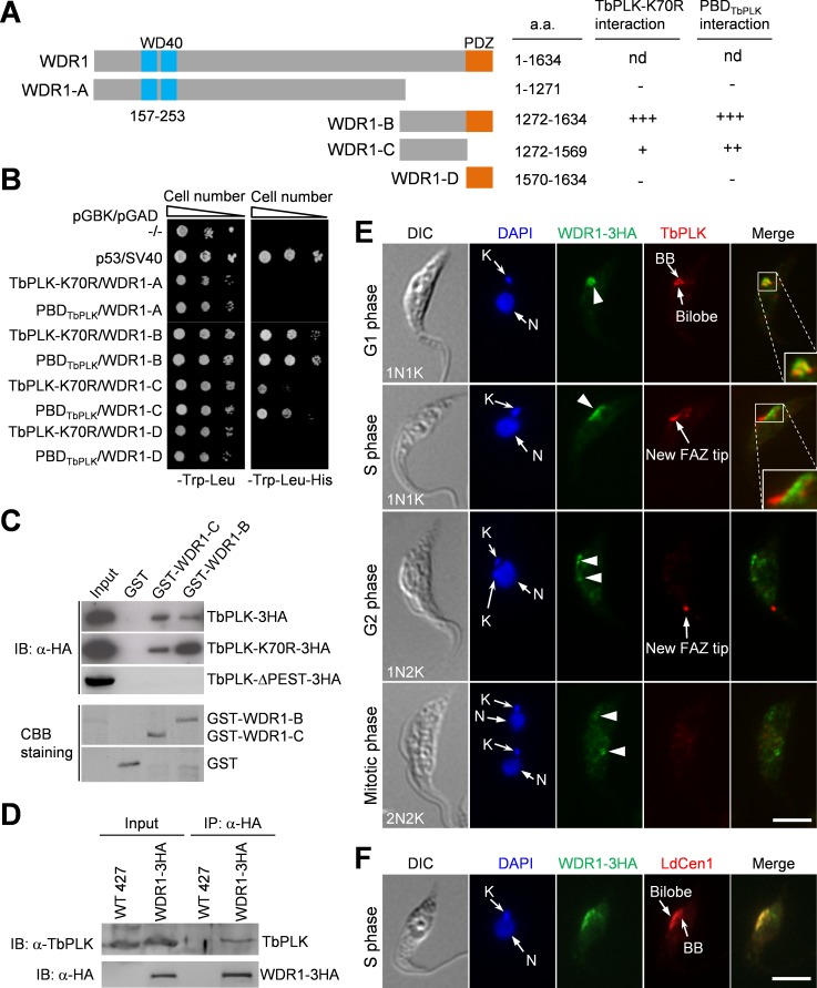
NCBITaxon:5691) · Huiqing Hu, Qing Zhou, Xianxian Han, Ziyin Li; CC BY, via PubMed Central · CC_BY_4_0 · source: PMC PMC5241021.1 ppat.1006146.g002 · DOI:10.1371/journal.ppat.1006146Synonyms: CRL4WDR1
Components
| Component | Type | Part of / acts on | Grounding | Genes | Stoichiometry | Essential | Role |
|---|---|---|---|---|---|---|---|
| Cullin4 scaffold subunit | Protein | part of | Reviewed label only (reviewed name; no accession asserted) | CUL4 | UNKNOWN | Defining cullin scaffold co-isolated with WDR1 and DDB1; direct contacts and copy number are unresolved.
| |
| DDB1 adaptor subunit | Protein | part of | Reviewed label only (reviewed name; no accession asserted) | DDB1 | UNKNOWN | Adaptor co-isolated with WDR1; a WDR1 N-terminal fragment retrieves DDB1 from cell lysate.
| |
| WDR1 substrate-recognition subunit | Protein | part of | Reviewed label only (reviewed name; no accession asserted) | WDR1 | UNKNOWN | Receptor-class constituent that associates with DDB1 through its N-terminal region and recognizes TbPLK through C-terminal fragments. Its depletion slows TbPLK turnover.
|
Canonical examples
NCBITaxon:5691Trypanosoma brucei — The paper studies procyclic cells: strain 29-13 for inducible RNAi and Lister 427 for endogenous tagging, with tagging also performed in RNAi lines. Two nonoverlapping WDR1 RNAi fragments and two clones per fragment were tested; the paper presents one representative clone after reporting similar phenotypes. The 927 locus identifiers are reference annotations, not the cultured strain. Yeast two-hybrid hosts and recombinant E. coli expression hosts are not canonical organisms for this complex. [DOI:10.1371/journal.ppat.1006146]
Functions
- regulation of TbPLK protein turnover — WDR1 depletion slows the loss of TbPLK during a cycloheximide chase and increases its abundance in procyclic T. brucei. This supports a turnover-regulatory role for the named association, but does not establish direct covalent ubiquitination of TbPLK by a purified complex.
DOI:10.1371/journal.ppat.1006146Figure 6A-C measures abundance at 24/48 hours of RNAi and a 0-6-hour chase after 24 hours of induction. Figure 6D detects less polyubiquitinated material in TbPLK immunoprecipitates, but the Results explicitly leave its identity unresolved: TbPLK itself and/or associated proteins.
- support of bilobe duplication and flagellum attachment — WDR1 RNAi impairs bilobe duplication, basal-body segregation and new flagellum-attachment-zone assembly, with detached flagella at 48 hours. These are perturbation-supported cellular outputs, not proof that every phenotype is mediated exclusively by direct TbPLK ubiquitination.
DOI:10.1371/journal.ppat.1006146Figures 4-5 show basal-body, bilobe, FAZ and attachment phenotypes after 48 hours of WDR1 RNAi. Figure 7 shows persistent phospho-Ser54 TbCentrin2 staining in late-cycle cells at 24 hours. TbPLK overexpression in S4 Figure phenocopies several effects, but is not a rescue or an epistasis test.
Mechanism graphs
Measured outputs of WDR1 depletion (FUNCTION)
Source-bounded protein and cellular readouts of a receptor-subunit intervention.
| Subject | Predicate | Object | Evidence |
|---|---|---|---|
| WDR1 RNAi induction | decreases abundance of | WDR1 substrate-recognition subunit |
|
| WDR1 RNAi induction | reduces | TbPLK degradation rate in chase |
|
| WDR1 RNAi induction | increases | whole-cell TbPLK abundance |
|
| WDR1 RNAi induction | decreases | polyubiquitinated material in TbPLK immunoprecipitate |
|
| WDR1 RNAi induction | yields cells retaining | late-cycle TbCentrin2 phospho-Ser54 staining |
|
| WDR1 RNAi induction | increases | flagellum detachment |
|
Discussions
- CURATION_TODO (RESOLVED): Separate CRL4WDR1 from generic Cul4 and other DDB1 complexes. Shared cullin or receptor names do not establish equivalence.
- CURATION_TODO (RESOLVED): Keep the WDR1-specific assembly distinct from Cullin-E and broader Cullin-D isolates. A scaffold identity is not the identity of all its receptor-specific complexes.
- CURATION_TODO (OPEN): Resolve exact component-family mappings and studied-strain protein examples. Broad families and reference-genome annotations do not supply exact experimental examples.
- CURATION_TODO (RESOLVED): Do not convert captured client proteins or experimental markers into obligatory constituents. Co-isolation and downstream readouts have different biological roles.
- CURATION_TODO (OPEN): Establish the complete catalytic assembly and direct covalent substrate reaction. Co-immunoprecipitation and chase data do not identify all catalytic subunits or ubiquitinated proteins.
- CURATION_TODO (OPEN): Resolve localization and architecture of the assembled CRL4WDR1 particle. Subunit immunofluorescence does not demonstrate colocalization of all constituents.
Evidence
DOI:10.1371/journal.ppat.1006146Hu, Zhou, Han and Li; PMID:28052114. Current PMC5241021.1 JATS identity, license and checksum verified. Complex-identification, depletion, turnover and overexpression Results, Discussion and relevant Methods inspected. Native Figures 2, 3 and 6 were visually inspected with media checksums verified. Supports the named association and measured intervention outputs; the Figure 8 causal model exceeds the direct biochemical resolution.DOI:10.1038/s41598-025-32077-9PMID:41413177; Table 1 maps the same Tb927.3.1290 scaffold to TbCul-D, not TbCul-E. The newer scaffold-isolation study identifies additional DCAF proteins and Rbx1, without resolving their membership in the WDR1-specific assembly. Used for nomenclature and boundary comparison only.
Curation history
2026-10-04T19:38:22Zcodex · CREATED_RECORD — Scaffolded by scripts/new_record.py2026-10-04T19:40:32Zcodex · CURATED_RECORD — Added three source-bounded constituent classes, verified broad Cul4 parent, procyclic Trypanosoma example, turnover and inheritance functions, nonmechanistic depletion graph, bounded native Figure 2 with verified CC BY 4.0 provenance, and explicit identity, catalytic, family and imaging limits.
Source: data/structures/other/crl4wdr1_ubiquitin_ligase_complex.yaml